
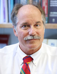
Affiliation : Professor Emeritus of Oncology and Previous Director Department of Flow & Image Cytometry at Roswell Park Comprehensive Cancer Center, USA
Title of the Talk/Lab :Monitoring &Tracking cell proliferation
Paul Wallace, Professor Emeritus of Oncology and Previous Director Department of Flow & Image Cytometry at Roswell Park Comprehensive Cancer Center, is recognized for his expertise in clinical flow cytometry with a strong background in immunology and research interests in antigen processing and presentation. He is Past President of the International Society for the Advancement of Cytometry (ISAC) a past Councilor of the International Clinical Cytometry Society (ICCS) and 2018 recipient of their Wallace H. Coulter award for lifetime achievement in clinical cytometry.Dr. Wallace Directed Roswell Park’s Flow and Image Cytometry for 19 years where he actively worked to build translational synergies between clinical and research flow cytometry. The clinical laboratory focused on the diagnosis and monitoring of patients with leukemia and lymphoma with an emphasis on minimal residual disease. Before joining Roswell Park, Dr. Wallace was an Assistant Professor of Immunology at Dartmouth Medical School, Lebaonon, NH (1993-2003); a cofounder of Zynaxis Cell Science, Inc., Malvern PA (1988-1991) the company that developed the PKH tracking dyes, and supervisor of Microbiology, Immunology, Serology, and Flow Cytometry at SmithKline Clinical Laboratories. He obtained his PhD from the Medical College of Pennsylvania in 1993 and his Masters from Idaho State University in 1979.
Lecture: Monitoring &Tracking cell proliferation: Cytometric definitions of “proliferation” vary, but the study of normal growth processes and how they go awry in tumors has been of interest to cytometrists since the inception of the field. Measurements of cell proliferation by flow cytometry can be broadly divided into two different categories – methods that measure aspects of DNA content or cell cycle and methods that track cell division by dye-dilution. Cell cycle analysis is a very common flow cytometric application, but its accurate measurement can test the limits of both technique and the flow cytometer. By using a DNA-specific stain, one can determine a DNA profile and thus determine the cell’s ploidy and percentage of cells in G0/G1, S, and G2/M. This information can be used to, for example, to monitor the aggressiveness of a tumor or the effect of an anticancer treatment. Measurement of the S-phase fraction based on DNA content tells us what proportion of cells in a sample are preparing for cell division at a given point in time. It does not, however, allow us to say how many divisions a given cell may have undergone in response to a stimulus, how much a particular cell subset may have expanded during that response or what fraction of a starting cell population went on to divide during the response. Flow cytometry can, however, be used to monitor the extent of cell division by: 1) staining cells with bright, stable, non-toxic fluorescent dyes that label bulk cell proteins or membranes; and 2) following the decrease in intensity (dye dilution) as the dyes are partitioned between daughter cells at each successive mitosis. The dye-dilution method has been used to monitor proliferation in vitro, to enumerate antigen specific cells, and to identify quiescent stem cells to name just a few applications. The principles of cell staining, analysis strategies, practical problems, and specific applications for each of the two major cytometric approaches will be covered.
Relevant Literature:
Please click the below button to access the registration form. Registration is open until
January 25th, 2022